The perennial fascination of ancient Egyptian mummies is being used by Queensland University of Technology researcher Dr Stephen Hughes to teach children about the application of physics to the field of archaeology.
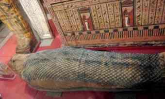 Dr Hughes has produced an education kit for teachers about his experience of "unwrapping" the secrets of a mummy of a young woman who died around 715 BCE and reconstructing her face from x-ray images. Using ordinary x-ray and CT x-ray scanning, the researchers could look inside the body without having to open the ornate, body-shaped coffin.
"The mummy was of a female of high rank called Tjetmutjengebtiu, or 'Jeni' for short, who was a singer in the great temple of Karnak, and had been held in the British Museum for 100 years," Dr Hughes said.
"I led a team of researchers using modern diagnostic technology to examine the mummy without leaving a pile of bones and dust, yards of linen and a broken coffin as usually happens when mummies are taken out of their coffin and unwrapped.
"CT scans enable the undisturbed internal arrangements of the mummy to be studied and for information about the bones to be gathered which would be difficult to do even if the mummy were unwrapped.
"Using the CT scan images we were able to produce a picture of Jeni's face . And we were able to see that her lungs, liver, stomach and intestines had been removed, embalmed, wrapped and placed back in the chest cavity. "
Dr Hughes said he had published several articles about his findings in academic journals but was asked by teachers to prepare some material for school-aged children.
"The teaching aids I have developed give children an introduction to how CT images are obtained and how they differ from x-rays while drawing them in to learn about how these technologies revealed information about 'Jeni's' life," he said.
"For example, Jeni's teeth were scanned in high resolution and the images generated a detailed reconstruction. The fact that her teeth were in good condition and the open shape of the roots of her wisdom teeth suggests she was between 19 and 23 when she died.
"By middle age, most ancient Egyptians' had lost several teeth, had abscesses and the enamel on the remaining teeth was worn down to pulp."
Dr Hughes said a 3D reconstruction of the skull showed that the embalmers had removed Jeni's brain through her nostrils and then stuffed the empty cranium with linen.
"The embalmers did this by putting a spike up the nose like a crochet hook, smashing through the ethmoid bone, which has the same consistency as a Crunchie chocolate bar, the mashing the brain and removing it bit by bit through the nose."
Dr Hughes said students had an almost universal interest in ancient Egypt and by using the example of the mummy they learnt about how physics contributed to knowledge of the real world, albeit from 2700 years ago.
He has spoken to the public and school groups on the topic and is available to talk to school students by contacting him on sw.hughes@qut.edu.au. This e-mail address is being protected from spam bots, you need JavaScript enabled to view it The education materials can be viewed at http://eprints.qut.edu.au/31522/
Source: Science Alert [July 16, 2010]